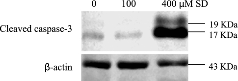
Figure 5.
Effects of SD on expression of cleaved caspase-3 in A431 cells. Cells were treated with SD (0–400 µM) for 48 hours, and then cells were collected by brief trypsinization. Total cell lysates were prepared and loaded to SDS-PAGE and Western blot analysis. Membranes were then probed with cleaved caspase-3 and β-actin antibodies followed by appropriate secondary antibody and ECL detection.

