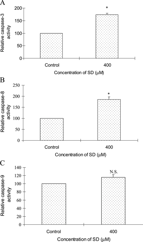
Figure 6.
Effects of SD on activities of caspase-3, -8, and -9 in A431 cells. Cells were treated with SD (0–400 µM) for 48 hours, and then enzymatic activity in cell lysates was quantified by measuring chromophores obtained from cleaved substrates. (A) Caspase-3 activity was assessed as the cleavage of DEVE-pNA to synthetic tetrapeptide DEVD and chromophore pNA. (B) Caspase-8 activity was determined by the cleavage of IETD-pNA to synthetic tetrapeptide IETD and chromophore pNA. (C) Caspase-9 activity was investigated by the cleavage of LEHD-pNA to synthetic tetrapeptide LEHD and chromophore pNA as detailed in the Materials and Methods section. Data are shown as the percentage of the absorbance values to controls and represent mean ± SE of three independent samples. *P < .05 indicates statistical significance in SD-treated groups compared with the control.

