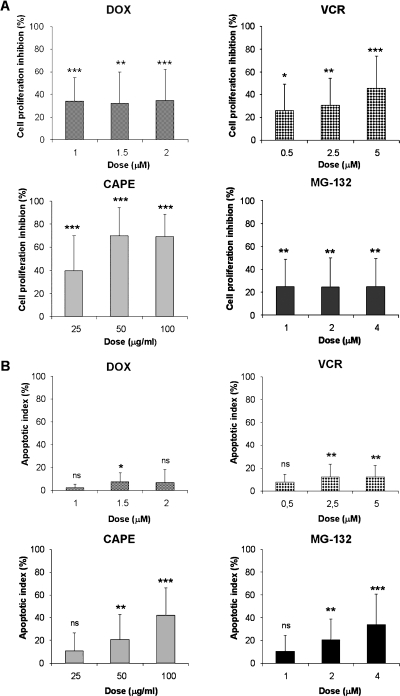
Figure 6.
CAPE and MG-132 treatment showed an antiproliferative and cytotoxic effect on PDL cells. (A) PDL cells were cultured in the presence of increasing concentrations of DOX, VCR, CAPE, or MG-132 for 24 hours. Results are presented as percentage of cell proliferation inhibition of each treatment. (B) Apoptosis induction evaluated after acridine orange and EtBr staining. PDL cells were treated for 24 hours with DOX, VCR, CAPE, or MG-132. Bars, mean ± SD of at least three independent experiments (ns indicates not significant, *P < .05, **P < .01, ***P < .001).

