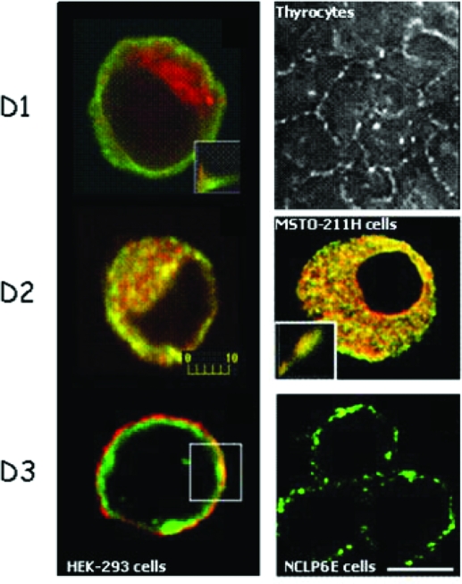
Figure 2.
Subcellular localization of deiodinases by immunofluorescence. HEK-293 cells were transiently transfected with FLAG-tagged D1, D2, or D3 (left). D1 (green) is located in the plasma membrane region and does not colocalize with the ER marker GRP78/BIP (red), in contrast to D2 (green), which is located in the ER. D3 (green) was colocalized to the plasma membrane with Na/K ATPase (red). Immunofluorescence staining of endogenously expressed deiodinases is shown in the right panel. D1 in porcine thyrocytes is found in the plasma membrane (image kindly provided by Dr. Peter Arvan, Ann Arbor, MI), whereas endogenously expressed D2 in MSTO-211H cells colocalized with GRP78/BIP in the ER. Endogenous D3 protein was detected in the plasma membrane of NCLP-6E cells. Scale bar, 10 μm. [Reprinted with permission from Baqui et al.: Endocrinology 141:4309–4312, 2000 (48), ©The Endocrine Society. Reproduced with permission of the Company of Biologists from Prabakaran et al.: J Cell Sci 112:1247–1256, 1999 (47); Curcio et al.: J Biol Chem 276:30183–30187, 2001 (49), ©ASBMB, Inc.; and Baqui et al.: J Biol Chem 278:1206–1211, 2003 (50), ©ASBMB, Inc.]

