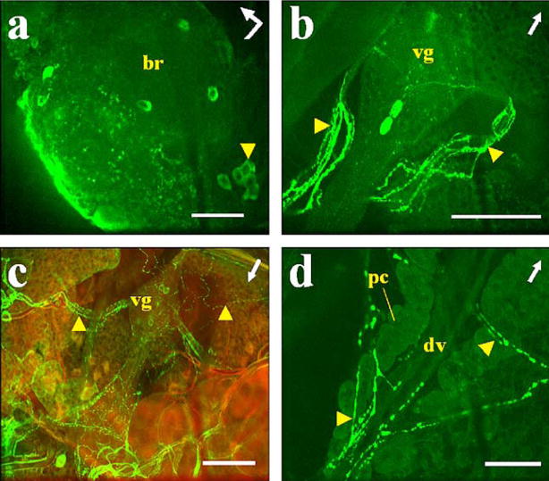
Fig. 2.
Immunostaining of AT in A. albimanus tissues. a Left protocerebral lobe of the brain (br) with a posterior group of labeled cells (arrowhead). Bent arrow Front (arrowhead) and left side (arrow end) of the brain. b, c Abdominal preparations with three cells labeled in ventral ganglia (vg) and varicosities labeled in ventral processes (arrowheads). c Combined images from confocal fluorescence (green) and light (red) microscopy. d Heart (arrowheads processes directly connected to the dorsal vessel containing immunostained varicosities, dv dorsal vessel of heart, pc pericardial cells). Arrows Anterior part of the mosquito. Bars 50 μm

