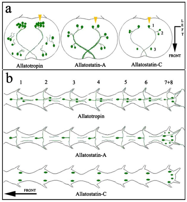
Fig. 6.
Representation of neuropeptide distribution in the central nervous system of A. aegypti and A. albimanus. The pattern of immunostaining for each of the three peptides is similar in both mosquito species. a Brains showing neurons and processes stained in the protocerebral lobes. Bent arrow Front (arrowhead) and left side (arrow end) of the brain (numbers relative location of cells). b Abdominal ganglia in the ventral nerve cord showing neurons and processes stained with the three different antibodies (numbers position of the ganglia). Arrow Anterior part of the mosquito

