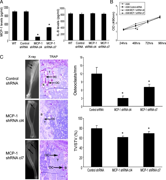
Figure 5.
Knockdown MCP-1 in A549 cells diminished the tumor growth in bone. (A) ELISA was performed in the CM from the selected A549 cells that were stably transfected with shRNA targeting of either MCP-1 or a scrambled control. *P < .001 compared with wild type (WT) or control shRNA cells. (B) MCP-1 knockdown in A549 cells minimally decreased the tumor cell proliferation as determined by MTS assay. *P < .01, significant difference from the control cells by t test. (C) Single-cell suspension of the selected A549 cells was injected into the right tibia of SCID mice. The tumor cells were allowed to grow for 4 weeks, at which time the mice were killed. Evidence of tumor-induced bone osteolysis was evaluated by x-ray, H&E staining, and TRAP staining. On the x-ray film, arrows point to the tumor-induced osteolysis. On TRAP staining, T represents tumor cells, B represents bone, OC represents osteoclast, and arrows point to the increased osteoclast activities. Osteoclast number per millimeter bone surface is determined by bone histomorphometry. Tumor volume versus non-bone soft tissue volume was quantified by bone histomorphometry. Results are reported as mean ± SD. *P < .001 compared with the control cells.

