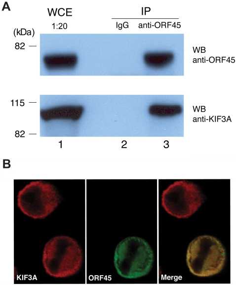
Figure 1. Interaction of KSHV ORF45 with KIF3A.
(A) BCBL-1 cells induced with TPA for 48 hours were lysed, and whole cell extracts (WCE) were immunoprecipitated (IP) with mouse IgG (negative control) or mouse monoclonal anti-ORF45 antibody. The immunoprecipitates were analysed by Western blotting (WB) with anti-ORF45 (upper panel) and anti-KIF3A antibodies (lower panel). Positions of the molecular mass standards (kDa) are shown toward the left. (B) BCBL-1 cells treated with TPA for 48 hours were fixed and permeabilized. Cells were subjected to double-labeled IFA using rabbit-polyclonal anti-KIF3A (red) and mouse monoclonal anti-ORF45 (green). The merged image is also shown.

