Figure 5.
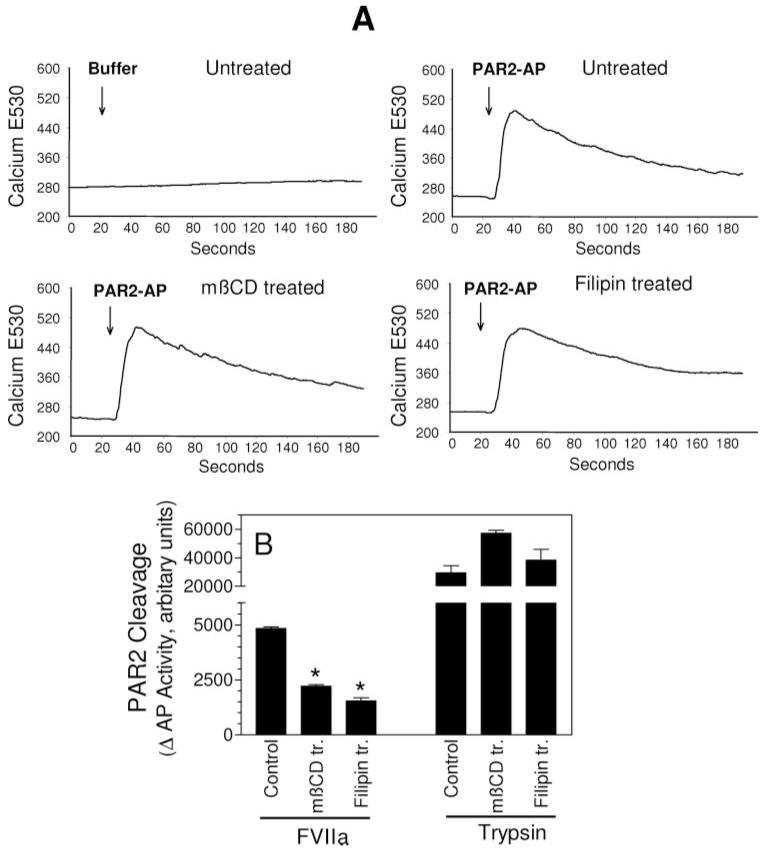
Membrane cholesterol depletion/modification specifically impairs TF-VIIa activation of PAR2. A, MDA231 cells cultured in glass-chambered slides were loaded with Fluo-4 and treated with mβCD (10 mmol/L for 1 hour) or filipin (5 μg/mL for 15 minutes). After mounting the slide on a microscope stage, live fluorescence images were obtained for 30 seconds, and then control vehicle or PAR2 agonist peptide (SLIGKV, 25 μmol/L) was added to the cells, and the imaging was continued for 3 minutes. Increase in intracellular Ca2+ levels were shown as increase in fluorescence at 530 nm emission wavelength. B, COS-7 cells transiently transfected with TF plus AP-PAR2 expression vectors were treated with a control vehicle, mβCD (10 mmol/L for 1 hour), or filipin (5 μg/mL for 15 minutes), and thereafter exposed to VIIa or trypsin (10 nM) for 1 hour, then soluble AP activity in the conditioned medium was measured. Results are expressed as AP activity in cells treated with VIIa or trypsin minus that in cells not treated with proteases, but subjected to mβCD and filipin treatments (n=3 ±SEM). *Significantly (P<0.05) differs from the control cells exposed to the same protease.

