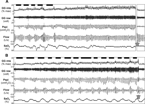
Figure 4.
Raw data for 10 min after sleep onset in a 45-year-old man with OSA (AHI = 57.1 events/h) who developed stable breathing (A) and a 49 year old male patient (AHI = 69.7 events/h) who did not develop stable breathing (B). Arterial oxygen saturation (SpO2), airflow (Flow), epiglottic pressure (PEPI), and both the raw and moving time averaged (MTA) genioglossus (GG) electromyogram are shown. Respiratory events marked by the technician are shown by black bars above the GG signal. Note that both individuals had increased GG activity as sleep progressed. However, only subject A was able to stabilize breathing (although still flow limited). Following full awakening (as shown by the arrow), genioglossus activity instantly fell to baseline in both subjects. In Subject A, 6.5 min of data are omitted prior to full awakening (broken axis), such that 10 min of data are shown for both individuals.

