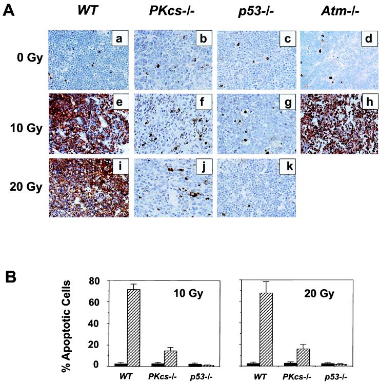
Figure 1.
Radiation-induced apoptosis in thymic tissues of wild-type (WT) , DNA-PKcs−/−, p53−/−, and Atm−/− mice. (A) Animals were mock-irradiated (a–d), irradiated with 10 Gy (e–h), or 20 Gy (i–k), and apoptosis was evaluated 10 hr postirradiation (e–k) by using the TUNEL in situ assay. (B) Graphic representation of IR-induced apoptosis in thymus. At least 3,000 cells were counted in a minimum of three animals for each genotype either before irradiation, or 10 hr after 10 Gy and 20 Gy, respectively. Each value represents the mean ± SD in percentage of apoptotic cells. Black bars represent values obtained in unirradiated animals, and hatched bars those from irradiated animals. Double staining with anti-CD34 antibody and TUNEL demonstrated that the apoptosis observed in DNA-PKcs−/− thymus represents mostly endothelial cells (data not shown).

