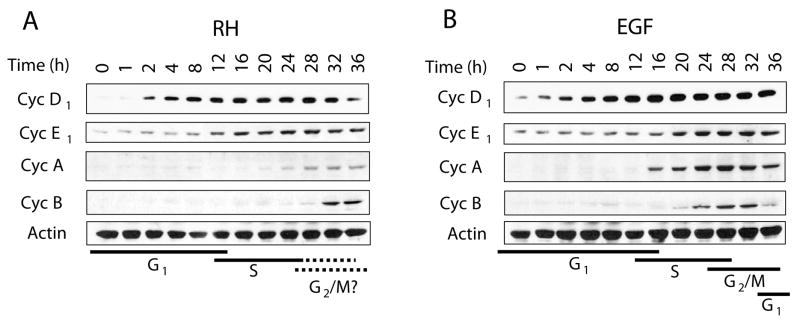
Fig. 4.
The cyclin profiles of infected cells correlate with entry into the cell cycle followed by interference in progression. Confluent HFF grown in 24-well plates were infected with T. gondii RH at an m.o.i. of 5:1 (A) or stimulated with 2 ng/ml of EGF (B) over a period of 36 h. Cells were processed for SDS-PAGE followed by immunoblotting using antibodies against cyclin D1, cyclin E1, cyclin A, cyclin B, and β-actin as a loading control. Both T. gondii infection and EGF treatment induced increases in the levels of cyclins D1 and E1 consistent with a G1/S transition. The kinetics of cyclins A and B on the other hand showed a delayed and abrogated response with infection. Results correspond to one representative experiment of three performed. Black lines below panels A and B represent the predicted stages of the cell cycle.

