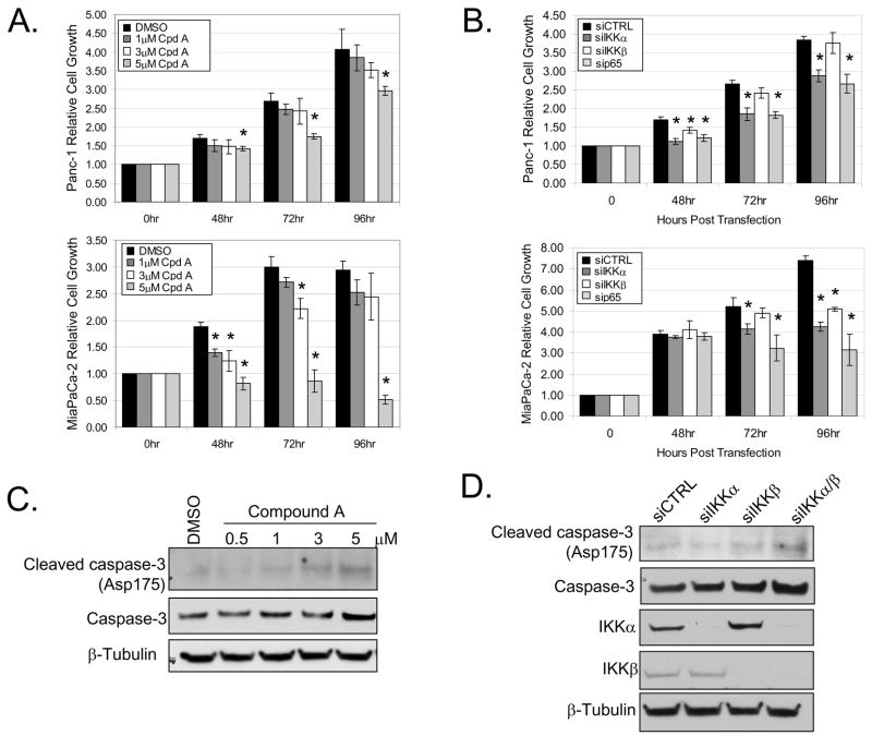
Figure 5. Blockade of IKK suppresses cell proliferation.
(A) Panc-1 and MiaPaCa-2 cells were treated with DMSO or 1, 3, and 5μM of IKKβ inhibitor (Compound A) for 48, 72, and 96 hours. Cell growth was measured in triplicate at each time-point using a colormetric MTS tetrazolium assay. (B) Panc-1 and MiaPaCa-2 cells were transiently transfected with 100nM siRNA targeted against IKKα, IKKβ, p65, and siCTRL. Cell growth was measured as described above at 48, 72, and 96 hours post-transfection. Data was normalized to the initial cell density prior to Compound A treatment or siRNA transfection respectively. Asterisks indicate statistical significance relative to DMSO or siCTRL (P < 0.05). (C) Panc-1 cells were treated with Compound A as described above for 24 hours. Whole-cell extracts were harvested and separated by SDS-PAGE. (D) Panc-1 cells were transiently transfected with 100nM siRNA targeted against IKKα, IKKβ, combined IKKα/β, and siCTRL. Whole-cell extracts were harvested, separated by SDS-PAGE, and immunoblotted using the specified antibodies.

