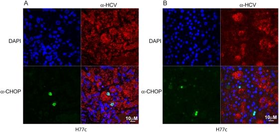
Figure 9. Confocal microscopy of HCV and CHOP/GADD153 in predominately infected or uninfected areas of HCV infected mice.
Panel (A) shows an area from an HCV infected liver that contains mostly infected cells. Panel (B) shows an area from an HCV infected liver that contains mostly uninfected cells. Liver sections from HCV H77c infected mice were stained using rabbit anti-CHOP and mouse anti-HCV antibodies, and the secondary antibodies were goat anti rabbit alexa 488 (green) and goat anti mouse poly-HRP, which was developed using tyramide-TMR (red). Nuclei were stained using DAPI (blue).

