
Figure 1.
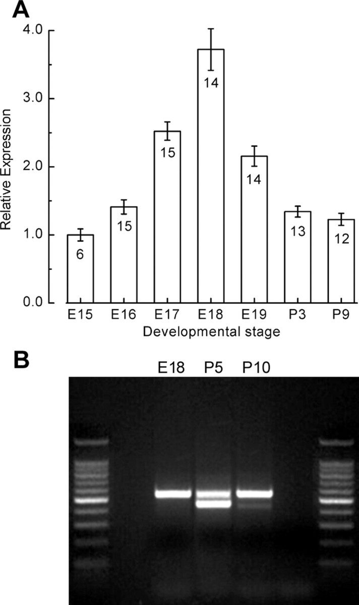
Expression of KCNQ4 mRNA in the mouse utricle. A, Quantitative real-time RT-PCR was used to examine the expression pattern of KCNQ4 in the developing mouse utricle. A standard curve was generated using tenfold dilution series of plasmid DNA that carried the coding sequence for mKCNQ4 was used to confirm primer efficiency. The relative expression levels are plotted as a function of developmental stage. The number of samples and SEs are shown for each stage. B, Image of an agarose gel electrophoresis showing the amplification products of an RT-PCR using primers that span exons 8 through 12 of mKCNQ4 and template harvested from the developmental stages indicated. A 1 kb ladder was run in lanes 1 and 5. Bands of 528 and 423 base pairs were amplified at P5 and P10.