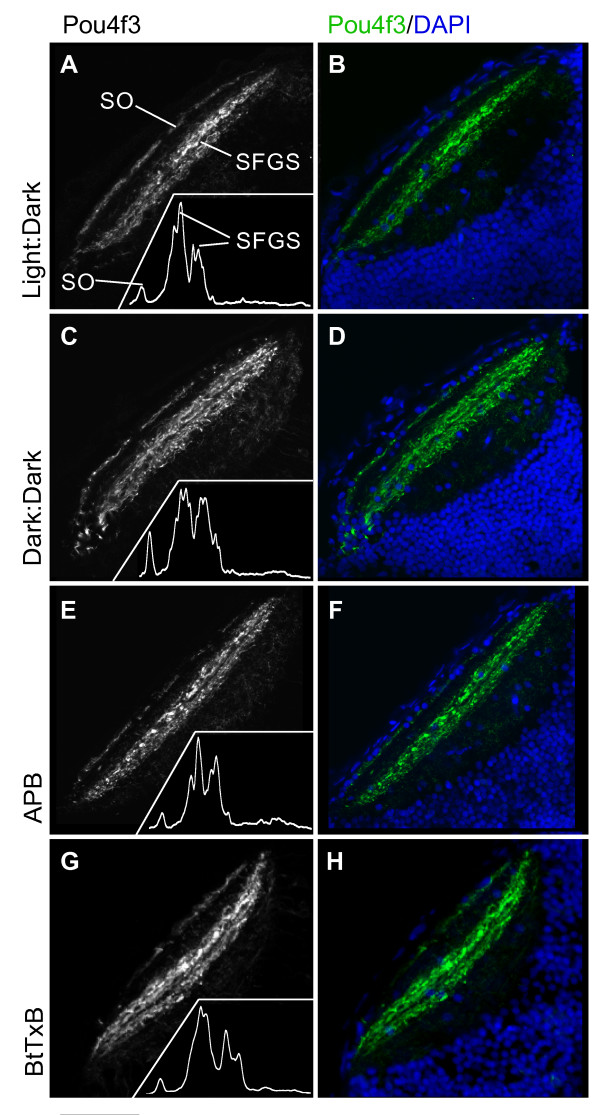
Figure 7.
Dark-reared, APB-treated, and BtTxB-injected larvae show proper GC axon targeting to tectal laminae. Horizontal sections of 5 dpf larval tecta showing Pou4f3:mGFP+ GC axons innervating the optic tectum, imaged by confocal microscopy. (A, C, E, G) Pou4f3:mGFP+ axons innervate the SO and two sublaminae of the SFGS. Insets: densitometric traces across the tectal neuropil, from superficial to deeper layers. (B, D, F, H) Same images of Pou4f3+ axons (green), with DAPI labeling (blue) to show the cell body and neuropil regions of the tectum. Scale bar 50 μm.

