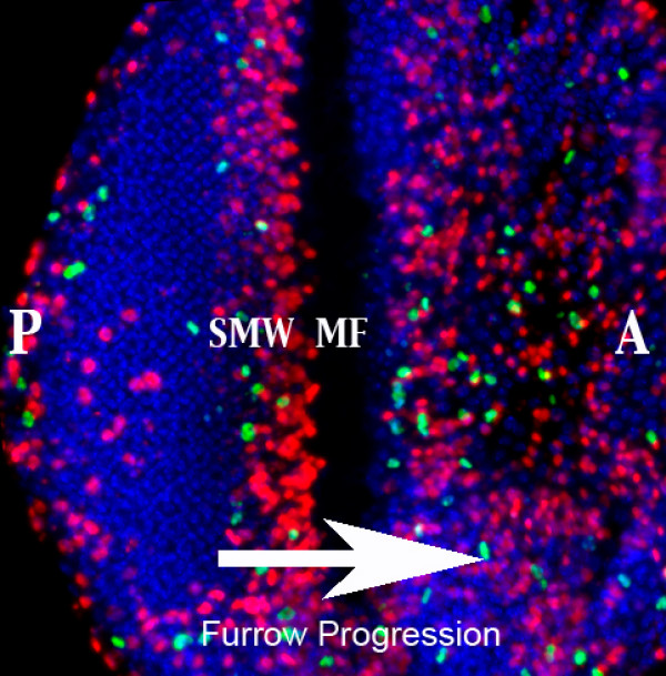
Figure 1.
Eye imaginal disc differentiation occurs in a wave that moves from posterior (P) to anterior (A). The margin between the asynchronously dividing anterior cells and the differentiated posterior cells is marked by the morphogenetic furrow (MF), where cells are delayed in G1. Mitotic division cycles become synchronized in the "Second Mitotic Wave" (SMW), which is composed of a tight band of DNA synthesis (marked by BrdU in red) and mitosis (marked by PH3 in green). The differentiated ommatidial clusters posterior of the furrow can be seen with the DNA stain in blue.

