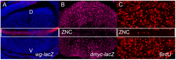
Figure 2.
Wg protein, dmyc expression and cell cycle patterning in the Drosophila wing pouch. (A) Wg protein (red) is strongly expressed along the dorsal-ventral boundary of the wing pouch. (B) α-gal antibody staining (pink) of dmyc-lacZ (w67c23P{lacW}l(1)G0354G0354; [109]) discs shows a pattern consistent with dmyc transcription throughout the cycling cells of the pouch and downregulation of dmyc within the G1 arrested cells of the ZNC. (C) The zone of non-proliferating cells (ZNC) can be seen by the reduced BrdU staining (red) for S-phase along the dorsal-ventral boundary. Wing imaginal discs are aligned dorsal (D) to the top of the image, ventral (V) the bottom.

