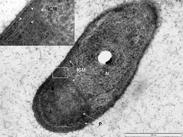
Figure 5.
Transmission electron micrograph of high-pressure frozen and cryosubstituted cell of Prosthecobacter dejongeii showing an intracytoplasmic membrane (ICM) surrounding a pirellulosome region containing a fibrillar nucleoid (N), paryphoplasm region at cell rim and a large invagination of rim paryphoplasm (P) at the cell pole. Inset: enlarged view of region of cell periphery showing continuity of the paryphoplasm at the cell rim with a large polar invagination of paryphoplasm, which is bounded by ICM which also defines an extension of the pirellulosome's riboplasm into the cell pole (see arrowheads). Bar – 500 nm.

