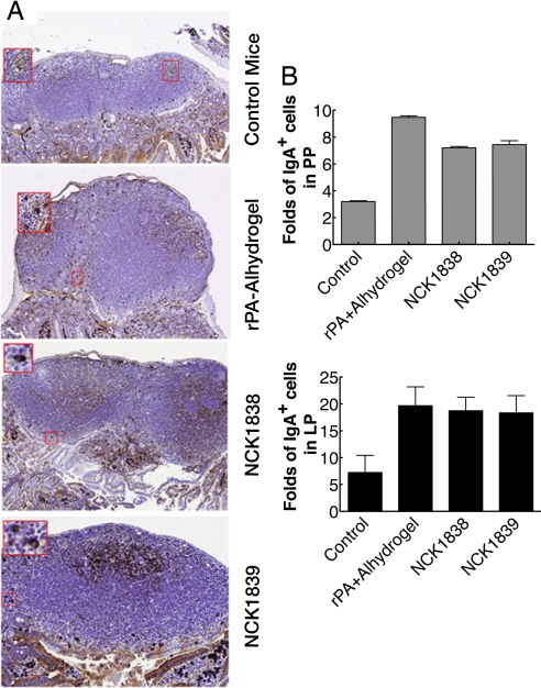
Fig. 5.
Detection of IgA-expressing cells within the small intestine. (A) After isolation of jejunum and ileum, these tissues were fixed in 10% formalin and processed into paraffin blocks. Serial tissue sections were then mounted on glass slides and IgA-expressing cells were detected with a rabbit anti-mouse IgA polyclonal and visualized with a goat anti-rabbit HRP secondary via scanning electron microscopy. Magnified areas are shown in red squares. (B) IgA+ cells of the lamina propria (LP) of villi and Peyer's patches (PP) were evaluated by a semiautomated quantitative image analysis system. Data are depicted as fold increases of IgA+-expressing plasma cells in LP and PP areas from all mice compared with unvaccinated controls. Data are representative of 3 independent experiments.

