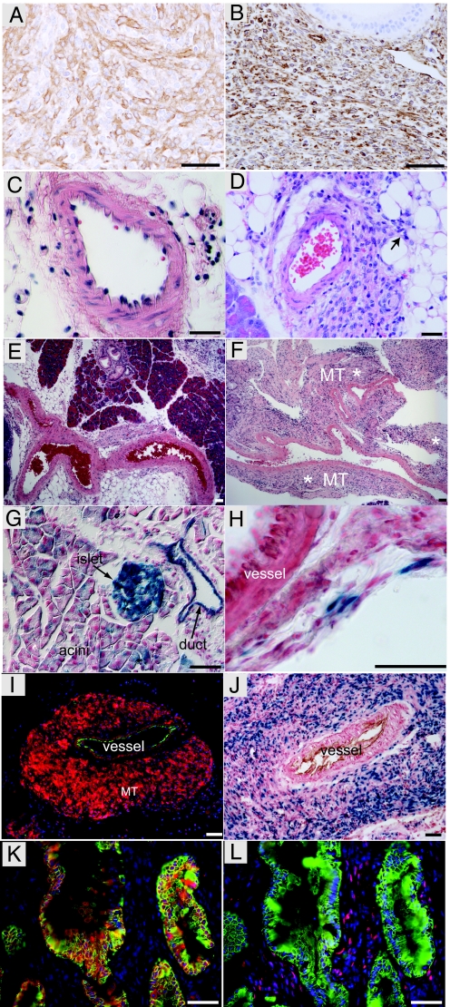
Fig. 2.
SmoM2 and KrasG12D cooperate to induce pancreatic mesenchymal tumors in PdxCre;KrasG12D;SmoM2 mice. (A) SMA expression by mesenchymal tumor in a PdxCre;KrasG12D;SmoM2 mouse. (B) Vimentin expression by mesenchymal tumor in a PdxCre;KrasG12D;SmoM2 mouse. (C) Muscular blood vessel in the pancreas of a control (PdxCre;KrasG12D) mouse at 4 weeks. (D) Small, mesenchymal tumor arising from a muscular blood vessel in the pancreas of a 4-week-old PdxCre;KrasG12D;SmoM2 mouse. Arrow highlights tumor extension into surrounding soft tissue. (E) Arteries in control PdxCre;KrasG12D animal at 8 weeks. (F) Artery and mesenchymal tumor in 8-week-old PdxCre;KrasG12D;SmoM2 mouse (MT* highlights tumor). (G) X-gal staining highlights the stochastic activity of PdxCre recombinase in pancreatic epithelium (duct, acini, and islet) when combined with the R26-LSL-LacZ reporter. (H) PdxCre activity is occasionally detected in adventitia surrounding blood vessels in PdxCre;R26R mice. (I) SmoM2-YFP expression (anti-YFP, red) in mesenchymal tumor (MT) arising from a muscular blood vessel (CD31, green) in a 12-week-old PdxCre;KrasG12D;SmoM2 mouse. (J) Hh signaling is active in mesenchymal tumor, as indicated by nuclear β-galactosidase enzymatic activity (blue) around blood vessel (CD31, brown). (K) SmoM2-YFP (red) expression in a higher-grade mPanIN epithelium (marked by CK19, green). (L) Hh signaling is not active in CK19-positive (green) neoplastic epithelium, as demonstrated by a lack of β-galactosidase immunoreactivity (red). β-galactosidase expression (red) in only observed in stromal cells adjacent to a higher-grade mPanIN (CK19, green; adjacent serial section to K). These images are representative of analysis of three animals per genotype. (Scale bars, 50 μm.)

