
Figure 1.
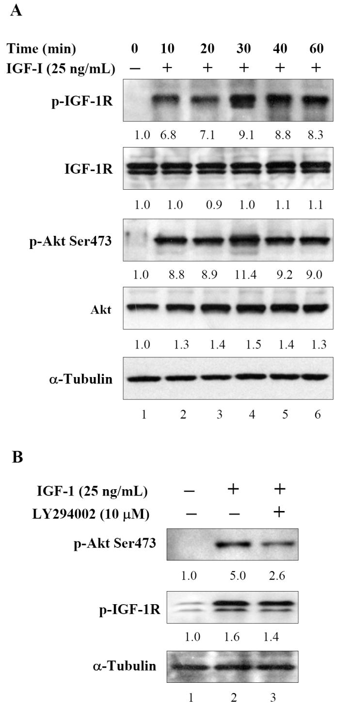
Effect of IGF-I on the phosphorylation of IGF-IR and downstream signaling in human prostate cancer DU145 cells. (A) The cells were serum starved and treated with 25ng/mL IGF-I and incubated for indicated times and later subjected to immunoblotting. An increase in IGF-IR and Akt phosphorylation was observed which attained highest levels at 30 min post IGF-I exposure. (B) DU145 cells were incubated with PI3K-Akt inhibitor LY294002 for 8 h prior to IGF-I stimulation. LY294002 treatment markedly dephosphorylated Akt whereas no significant effect was observed on IGF-IR phosphorylation. The blots were stripped and reprobed with anti-α-tubulin antibody to ensure equal protein loading. Fold change represents the protein level of the IGF-I treated cells relative to the control cells treated with vehicle and the resulting protein levels were then normalized to the α-tubulin protein. Details are described in ‘Materials and Methods’.