
Figure 4.
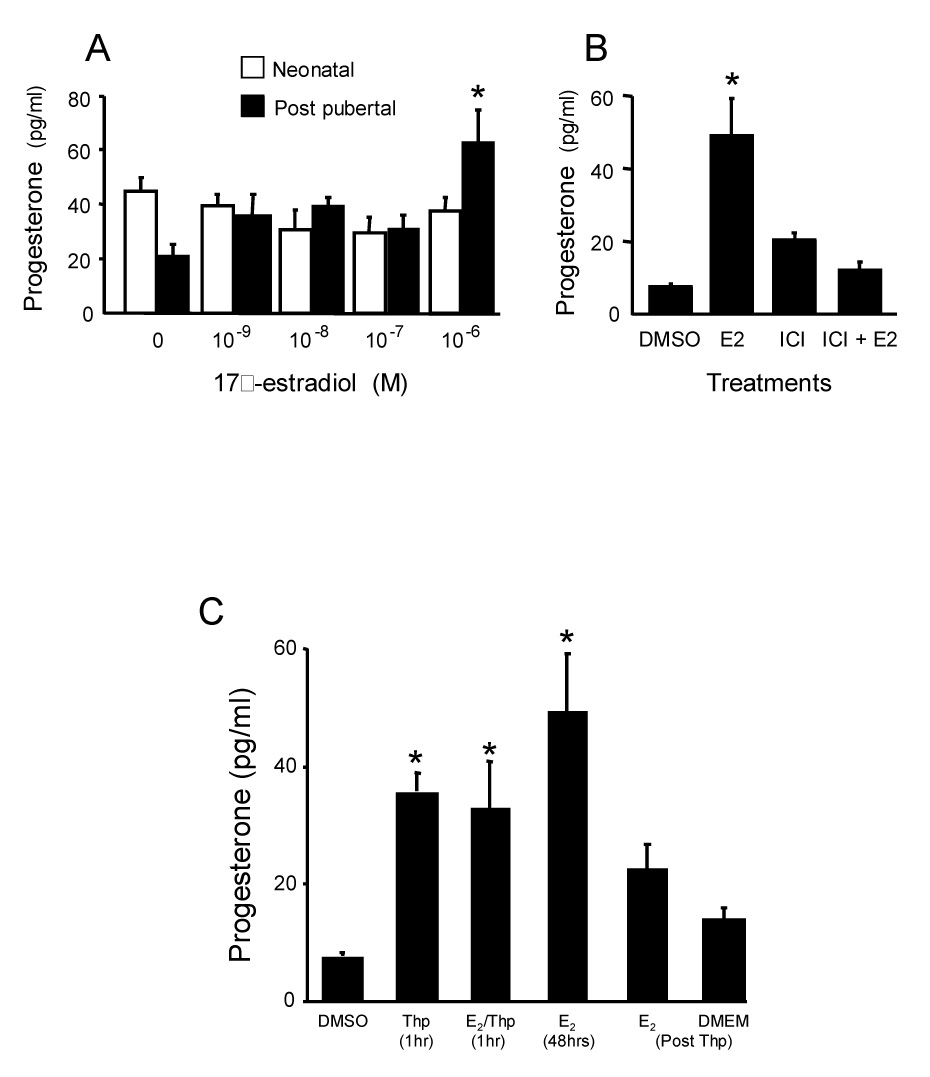
A. Effect of estradiol treatment of neonatal and post-pubertal hypothalamic astrocytes in vitro. The cells were steroid-starved for 24 hours and then incubated for 48 hours with indicated concentrations of 17β-estradiol or steroid-free media (0). Levels of progesterone in the supernatants were measured by radioimmunoassay. The neonatal astrocytes did not increase progesterone levels in the media in response to any estradiol dose tested. Post-pubertal astrocytes increased progesterone levels after treatment with estradiol. This increase was statistically significant at 10−6 M 17β-estradiol. Values are reported as mean ± SEM (n=8). * indicates significantly greater within developmental age group compared with 17β-estradiol free group (0) p < 0.05 (SNK). Figure 4B. Antagonism of 17β-estradiol (E2; 10−6 M) induction of progesterone levels in media from primary post-pubertal hypothalamic astrocyte cultures from female rats by blocking estrogen receptors. Steroid-starved astrocytes were incubated for 48 hours with either E2-free media with DMSO (DMSO), the vehicle used to dissolve the 10−6 M ICI 182,780 (ICI), an estrogen receptor antagonist, E2, ICI, or ICI + E2. Levels of progesterone in the supernatants were measured by radioimmunoassay. Data are means ± SEM (n = 4). * indicates values significantly greater than all other treatment groups (p < 0.05, SNK). Figure 4C. Effect of thapsigargin induced release of internal stores of Ca2+ on progesterone synthesis in astrocyte cultures obtained from post-pubertal female rats. Astrocytes were treated with thapsigargin (Thp) or Thp (10−7 M) supplemented with 10−6 M estradiol (E2/Thp) for one hour. The media was collected and replaced with either estradiol-free DMEM/F12 (DMEM Post Thp) or 10−6 M estradiol (E2 48hrs Post Thp). The progesterone concentration in the medium significantly increased following treatment with either E2 for 48 hr, Thp for one hr or Thp+ E2 for one hr. When the media was replaced with DMEM/F12 (DMEM) following Thp treatment there was no increase of progesterone concentration above baseline. Following one hour Thp treatment, subsequent exposure to E2 for 48 hr did not statistically increase the concentration of progesterone in the supernatant. Data are mean ± SEM (n = 4). * = indicates values significantly greater than control media, DMEM + DMSO (DMSO; p < 0.05 SNK; modified from (Micevych et al., 2007).