Figure 2.
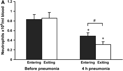
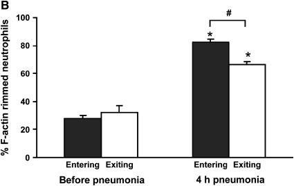
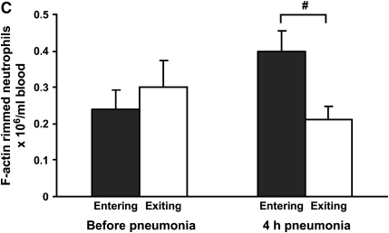
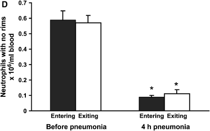
(A) Neutrophil counts in blood samples drawn simultaneously from the venous (entering the lungs) and arterial (exiting the lungs) circulation. Venous and arterial neutrophil counts were similar before pneumonia. Four hours after initiation of S. pneumoniae pneumonia, both venous and arterial counts were less than before pneumonia. The count was less in blood exiting the lungs compared with that entering the lungs, indicating that neutrophil sequestration within the lungs was occurring. (B) Percentage of circulating neutrophils that contained F-actin rims in S. pneumoniae pneumonia. The percentage of circulating neutrophils with F-actin rims increased by 4 h after instillation of organisms. At 4 h, the percentage of F-actin–rimmed neutrophils was less in venous blood than in arterial blood. (C) Number of F-actin–rimmed neutrophils in venous and arterial blood samples before and during S. pneumoniae pneumonia. The numbers of F-actin–rimmed neutrophils in venous blood and arterial blood were similar before pneumonia. Four hours after initiation of pneumonias, fewer F-actin–rimmed neutrophils were present in the blood exiting than entering the lungs, indicating that neutrophils containing F-actin rims sequestered in the lungs. (D) Number of neutrophils without F-actin rims in S. pneumoniae pneumonia. Neutrophils without rims were similar in number in venous and arterial blood at both time points. *Significantly less than the value determined at the same site before pneumonia, p < 0.05. #Significantly less than the value in the venous blood entering the lungs, p < 0.05.


