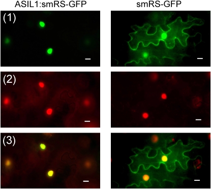
Figure 2.
Nuclear localization of ASIL1 Fused to smRS-GFP in Tobacco Epidermal Cells.
Plasmids that carried either construct p35S∷ASIL1:smRS-GFP or negative control p35S∷smRS-GFP were introduced into tobacco leaf cells by infiltration. All leaf tissues were stained with 4',6-diamidino-2-phenylindole (DAPI) and viewed using fluorescence microscopy with blue excitation to detect GFP fluorescence and UV excitation to detect DAPI. GFP is shown in green (1) and DAPI is shown in red (2). Merged images are shown at the bottom (3). Bars = 10 μm.

