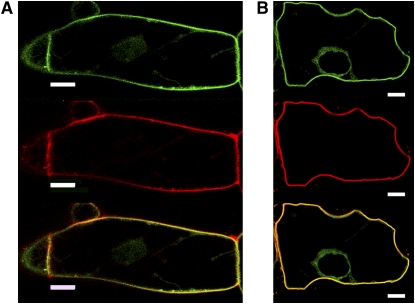
Figure 2.
Localization of GFP-BD-CVIL to the Plasma Membrane in BY-2 Cells.
GFP-BD-CVIL expression was induced for 15 h with 10 μM dexamethasone before treatment with the fluorescent marker FM4-64 (6.7 μg/mL). The green channel was set for detection of GFP (top panel) and the red channel for detection of the PM marker FM4-64 (middle panel). Merged images are shown in yellow and indicate colocalization of GFP-BD-CVIL and FM4-64 (bottom panel). Bars = 10 μm.
(A) In the absence of 0.23 M d-mannitol.
(B) In the presence of 0.23 M d-mannitol, which induces plasmolysis. The images show single optical sections through the center of the cell, including the nucleus.

