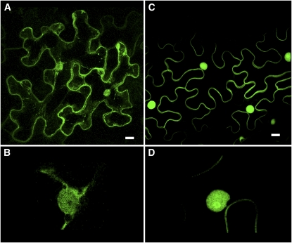
Figure 7.
Localization of GFP-CaM61 and GFP-BD-CVIL in Epidermal Cells of N. benthamiana Leaves.
(A) Localization of GFP-CaM61, showing GFP fluorescence associated with the cell periphery, nucleus, and cytoplasmic strands.
(B) At higher magnification, GFP-CaM61 is observed in the nucleus, but not the nucleolus.
(C) Localization of GFP-BD-CVIL, showing GFP fluorescence associated with the plasma membrane and nucleus, but not cytoplasmic strands.
(D) At higher magnification (threefold electronic zoom), GFP-BD-CVIL is observed to be concentrated in the nucleolus.
Bars = 10 μm. [See online article for color version of this figure.]

