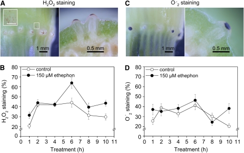
Figure 1.
H2O2 and O2.− Are Present in Epidermal Cells that Undergo Cell Death.
(A) Third node and cross section through a third node of a rice cv PG56 stem section stained with DAB to visualize H2O2. Stem sections were treated with 150 μM ethephon for 6 h. The inset shows hair-like structures on the epidermis that do not cover a root primordium (white rectangle). The bar in the inset = 0.5 mm.
(B) Percentage of epidermal patches above adventitious roots that showed H2O2 staining after treatment of stem sections with 150 μM ethephon for up to 10 h. DAB and ethephon were both applied at time 0 h. Results are averages (±se) from 19 to 48 stem sections analyzed per treatment. Each node contains 15 to 20 adventitious root primordia. The rate induced with ethephon at 6 h was significantly different from others at P < 0.001 (Tukey test).
(C) Third node and cross section through a third node of a rice cv PG56 stem section stained with NBT to visualize O2.−. Stem sections were treated with 150 μM ethephon for 6 h.
(D) Percentage of epidermal patches above adventitious roots that showed O2.− staining after treatment of stem sections with 150 μM ethephon for up to 10 h. NBT and ethephon were both applied at time 0 h. Results are averages (±se) from 32 to 49 stem sections analyzed per treatment. Rates are not significantly different at P < 0.001 (Tukey test).

