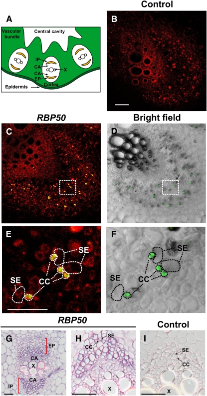
Figure 3.
In Situ RT-PCR– and RNA in Situ Hybridization–Based Detection of RBP50 Transcripts in Companion Cells.
(A) Schematic transverse section of a portion of the pumpkin petiole/stem. Vascular bundles are composed of internal and external phloem (IP and EP, respectively) and xylem (X), with an intervening cambium (CA).
(B) to (F) Transverse sections from pumpkin stem tissue analyzed by in situ RT-PCR using RBP50 gene-specific primers. Images were collected with a confocal laser scanning microscope; positive signal (green) represents incorporation of Alexa Fluor–labeled nucleotides.
(B) Negative control in which primers were omitted from the RT-PCR mixture. Red fluorescence represents tissue autofluorescence. Bar = 500 μm (common for [B] to [D]).
(C) Transverse section of a vascular bundle demonstrating the presence of RBP50 in phloem cells. Note that the green signal from the Alexa Fluor–labeled nucleotides appears yellow due to the red autofluorescence background.
(D) Bright-field image of (C) used to illustrate the cellular architecture of the vascular bundle. RBP50 transcripts (green fluorescent signal) accumulated in companion cells.
(E) Higher magnification of the boxed area shown in (C). CC, companion cell; SE, sieve element. Bar = 100 μm (common for [E] and [F]).
(F) Higher magnification of the boxed area shown in (D). Images presented are representative of those obtained from at least three replicate experiments.
(G) to (I) Pumpkin stem sections analyzed by in situ hybridization. Transverse sections were hybridized with an in vitro–transcribed antisense RNA probe to RBP50 ([G] and [H]); purple signal represents the presence of RBP50 transcripts in the small companion cells. Control in situ hybridization was performed with an in vitro–transcribed sense RNA probe to RBP50 (I). Bars = 500 μm.

