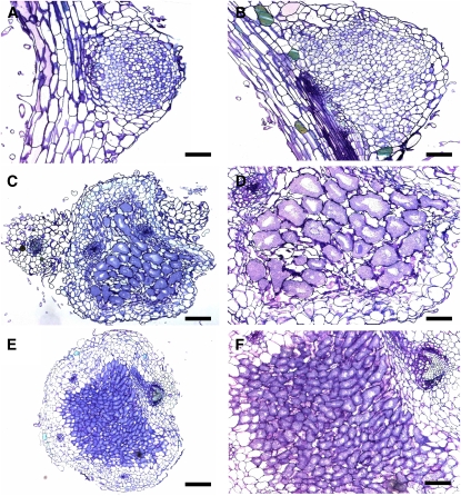
Figure 2.
Light Microscopy of Nodule Sections.
(A) Thin section of pir1-1 noninfected white bump.
(B) Close-up of nap1-1 noninfected bump.
(C) Thin section of large, red, infected nap1-1 nodule.
(D) Close-up of bacteroid-containing cells in large, red nap1-1 nodule. Note the enlarged size of the cells.
(E) Wild-type nodule.
(F) Wild-type nodule; close-up of the bacteroid-containing cells.
Bars = 100 μm in (A), (C), and (E) and 50 μm in (B), (D), and (F). [See online article for color version of this figure.]

