Figure 1.
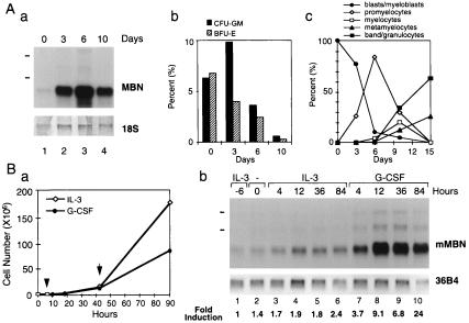
MBN is a G-CSF-responsive gene. (A) Expression of MBN during growth and differentiation of CD34+ cells toward the myeloid lineage. (a) Autoradiogram of MBN mRNA expression in CD34+ cells cultured in the presence of G-CSF. Total RNA (1.8 μg) was loaded in each lane. Positions of the 28S and 18S rRNAs are indicated on the left as size markers; Lower is methylene blue-stained 18S rRNA on membrane after transfer as assessment of RNA quantities in each lane. Days of culture are as indicated. (b) In vitro colonies formation. Cells harvested at 0, 3, 6, and 10 days were seeded in semi-solid culture conditions and percent of colony-forming unit–granulocyte/monocyte and burst forming-unit–erythroid cells were assessed at day 12. (c) Morphological differentiation of CD34+ toward the myeloid lineage. (B) Induction of MBN expression by G-CSF in Ba/F3/G-CSFR cells. (a) Growth of Ba/F3/G-CSFR cells with IL-3 or G-CSF. Arrowhead indicates addition of either IL-3 or G-CSF. Arrow indicates that cells were split at 1 × 10⁶ cells per ml. Concentrations of IL-3 and G-CSF were maintained at 0.1 ng/ml and 1 ng/ml, respectively. (b) Autoradiogram of murine MBN (mMBN) mRNA expression in Ba/F3/G-CSFR cells cultured in IL-3- or G-CSF-containing medium. Ba/F3/G-CSFR cells maintained in IL-3-containing medium (lane 1) were washed with factor-free medium. After 6 h incubation in factor-free medium (lane 2), cells were cultured with either IL-3 (lanes 3–6) or G-CSF (lanes 7–10) for the indicated times. Total RNA (2 μg) was loaded in each lane. Positions of the 28S and 18S rRNA are indicated on the left as size markers. Lower is an autoradiogram of 36B4 mRNA expression as assessment of RNA quantities in each lane. Fold inductions were calculated by the ratio of mMBN expression to 36B4 expression.

