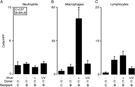
Figure 2.
Transplantation and respiratory viral infection combine to selectively enhance macrophage accumulation. In (A–C), transplantation conditions were as described in Figure 1A. Fourteen days post-transplantation, grafts were harvested and representative sections underwent immunolabeling with rat anti-mouse neutrophil IgG to identify neutrophils (A), rat anti–Mac-3 to identify macrophages (B), or rat anti-CD3 to identify lymphocytes (C). For each condition nonimmune IgG control gave no signal above background (data not shown). Immunolabeled cells in the lumen of the graft from each condition underwent quantitation (cells per HPF, ×400). Values represent means ± SEM (n = 4 or 5) from representative slides. *p < 0.05 compared with C → C and C → B.

