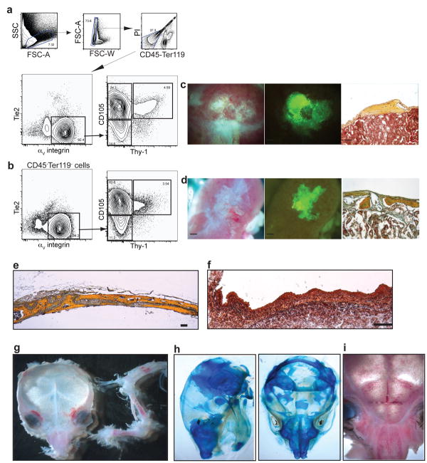
Figure 4.
Skeletal progenitors from mandible and calvaria do not form HSC niches efficiently. a-b, Representative FACS profile of homogenized 15.5 dpc mandible (n=6) (a) or calvaria (n=6) (b) pre-gated on live CD45-Ter119- cells. 2000 sorted GFP+ CD105+Thy-1-. c-d, Cells from mandible (c) or calvaria (d) were transplanted under renal capsule and harvested after 32 days. e, Pentachrome stained cross section of mouse parietal bone at 4 weeks. f, Pentachrome stained cross section of equivalent area in e15 dpc fetal calvaria. g-i, Marrow pockets in calvaria are concentrated in facial areas corresponding to cartilaginous regions (n=3). Limb bones are juxtaposed to skull for comparison. h, Dorsal and lateral views of newborn calvaria show cartilaginous regions that stain with alcian blue. (Scale bar in brightfield and GFP images = 500 μm, in pentachrome images = 100 μm, in (f) =25 μm).

