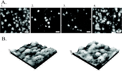
FIG. 4.
(A) Two-dimensional confocal images after 16 h of biofilm growth. Panel 1, S. oneidensis WT; panel 2, DL13; panel 3, DL13 supplemented with 5 μM AI-2; panel 4, DL13 pluxS. The scale bars represent 100 μm. (B) Three-dimensional projections of confocal images after 48 h of growth. Left, the S. oneidensis WT; right, DL13. Both three-dimensional projections have the dimensions of 900 μm by 900 μm by 60 μm. All images were taken with a 10× objective lens.

