FIG. 1.
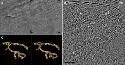
Cryoelectron tomography of a freeze-hydrated section of G. obscuriglobus. (A) A 15-nm-thick tomographic slice from the reconstructed volume shows the intracytoplasmic meshwork of double membranes. (B) The region framed in panel A shows the plasma membrane (PM) and the morphology of the intracytoplasmic membrane (ICM) system. The membrane curvature and junctions are indicated by arrowheads. (C) Stereo image of the membranes found within the tomogram shown in panel B. The surface-rendering view shows that the internal membranes in G. obscuriglobus are continuous with the ICM. Scale bars, 250 nm (A) and 125 nm (B).

