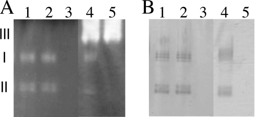
FIG. 1.
Detection of catalase and peroxidase activities in extracts from M. loti wild-type and mutant cells. Extracts from cells grown in TY medium to an OD600 of 0.3 were electrophoresed through nondenaturing polyacrylamide gels. (A) Catalase activity was negatively visualized as colorless bands (I, II, and III) on an oxidized-DAB background due to H2O2 consumption by electrophoresed catalase in the presence of externally added horseradish peroxidase and H2O2. (B) Peroxidase activity was visualized as dark bands of oxidized DAB in the presence of H2O2, and 30 μg of protein was loaded per lane. The other conditions are described in Materials and Methods. The extracts were from MAFF303099 (lanes 1), ML2101D (lanes 2), ML6940DS (lanes 3), MAFF303099 harboring p2101EX (lanes 4), and ML6940DS harboring p2101EX (lanes 5).

