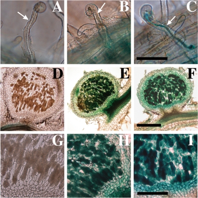
FIG. 7.
Histochemical detection of catalase gene expression during M. loti-L. japonicus nodule development. β-Galactosidase activity was detected using X-Gal as a substrate. The photographs depict MAFF303099 (A, D, and G), MLKEZ01 (katE-lacZYA) (B, E, and H), and MLKGZ01 (katG-lacZYA) (C, F, and I). (A to C) Root hairs. (D to I) Sections of 7-week-old nodules. Scale bars = 100 μm (A to C), 200 μm (G to I), and 500 μm (D to F). The arrows indicate infection threads.

