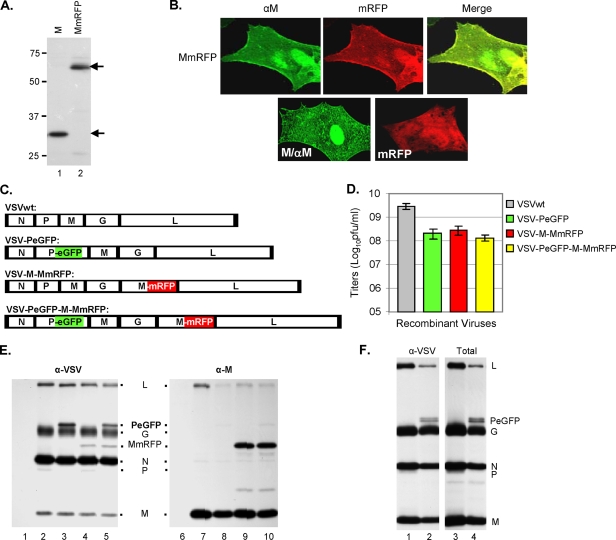
FIG. 1.
Recovery and characterization of recombinant VSV encoding fluorescent fusion proteins. (A) Expression of M and MmRFP fusion proteins. BHK-21 cells infected with vTF7-3 and transfected with a plasmid encoding M (lane 1) or MmRFP (lane 2) were radiolabeled with Expre35S35S label for 2 h at 16 hpt. The radiolabeled proteins were immunoprecipitated with anti-VSV antibody, separated by SDS-PAGE, and detected by fluorography. Size markers in kDa are shown on the left. The M and MmRFP proteins are identified with arrows on the right. (B) Epifluorescence and immunofluorescence staining of cells expressing the MmRFP, M, or mRFP protein. (C) Recombinant VSV genome plasmids. VSVwt, the wt VSV genome, with the N, P, M, G, and L genes shown in rectangular boxes; VSV-PeGFP, genome encoding PeGFP in place of wt P; VSV-M-MmRFP, genome encoding MmRFP at the G-L gene junction; VSV-PeGFP-M-MmRFP, genome encoding MmRFP at the G-L gene junction of the VSV-PeGFP genome. Intergenic regions as well as 3′-leader-gene and 5′-trailer sequences are shown in black boxes. (D) Growth of recombinant viruses. BHK-21 cells were infected with various recombinant VSVs at an MOI of 5. Growth of each recombinant virus was determined by plaque assay of culture supernatants at 16 hpi. Virus titers shown are averages from three independent experiments, with the standard deviations indicated by error bars. (E) Analysis of viral proteins in cells infected with recombinant VSVs. BHK-21 cells were infected with wt VSV (lanes 2 and 7), VSV-PeGFP (lanes 3 and 8), VSV-M-MmRFP (lanes 4 and 9), or VSV-PeGFP-M-MmRFP (lanes 5 and 10) at an MOI of 10 or were left uninfected (lanes 1 and 6). The proteins were radiolabeled for 1 h at 4 hpi, immunoprecipitated with anti-VSV antibody (lanes 1 to 5) or with anti-M antibody (lanes 6 to 10), separated by SDS-PAGE, and detected by fluorography. The viral proteins along with the fusion proteins are identified in the middle. (F) Examination of proteins incorporated into extracellular virions. The viral proteins in cells infected with VSV or VSV-PeGFP-M-MmRFP were radiolabeled with Expre35S35S label for 12 h at 4 hpi, and the proteins incorporated into the purified virions (VSV, lanes 1 and 3; and VSV-PeGFP-M-MmRFP, lanes 2 and 4) were examined by immunoprecipitation with anti-VSV antibody (lanes 1 and 2) or as total proteins (lanes 3 and 4). The positions of the viral proteins are shown on the right.

