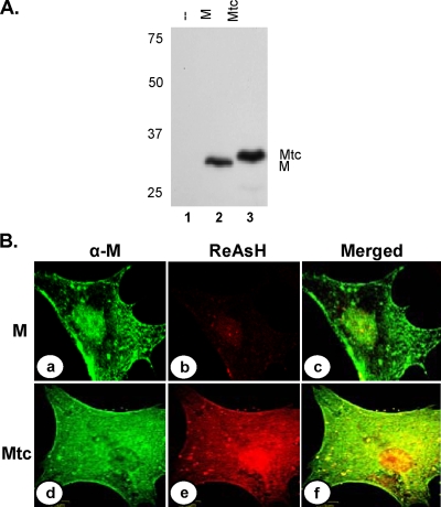
FIG. 2.
Expression of Mtc fusion protein in transfected cells. (A) Cells transfected with a plasmid encoding M (lane 2) or Mtc (lane 3) protein or mock-transfected cells (lane 1) were radiolabeled with Expre35S35S label for 2 h at 16 hpt. The radiolabeled proteins were immunoprecipitated with anti-VSV antibody, analyzed by SDS-PAGE, and detected by fluorography. Size markers in kDa are shown on the left. The M and Mtc proteins are identified on the right. (B) Biarsenical dye ReAsH labeling of cells expressing the M (a, b, and c) or the Mtc (d, e, and f) protein. Transfected cells were treated with ReAsH as described in Materials and Methods. Following washing of the dye, the cells were fixed and subjected to immunofluorescence staining with anti-M antibody and anti-mouse Alexa 488-conjugated secondary antibody. ReAsH staining (b and e), anti-M staining (a and d), and merged images of staining of the same cells (c and f) are shown.

