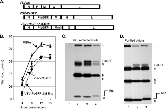
FIG. 3.
Recovery of VSV encoding Mtc. (A) Genome organization of wt VSV, VSV-PeGFP, and VSV-PeGFP-ΔM-Mtc. (B) Single-step growth kinetics of recombinant viruses. Virus titers shown are averages from three independent experiments, with the standard deviations indicated by error bars. (C) Analysis of viral proteins in cells infected with recombinant VSVs. BHK-21 cells were mock infected (lane 1) or infected with wt VSV (lane 2), VSV-PeGFP (lane 3), or VSV-PeGFP-ΔM-Mtc (lane 4) at an MOI of 10. The proteins were radiolabeled for 1 h at 4 hpi, immunoprecipitated with anti-VSV antibody, separated by SDS-PAGE, and detected by fluorography. The viral proteins along with the fusion proteins are identified on the right. (D) Examination of proteins incorporated into purified virions. Viral proteins in cells infected with VSV, VSV-PeGFP, or VSV-PeGFP-ΔM-Mtc were radiolabeled as described in the legend to Fig. 1F, and the proteins incorporated into purified virions (VSV, lane 1; VSV-PeGFP, lane 2; and VSV-PeGFP-ΔM-Mtc, lane 3) were examined. The positions of various proteins are shown on the right.

