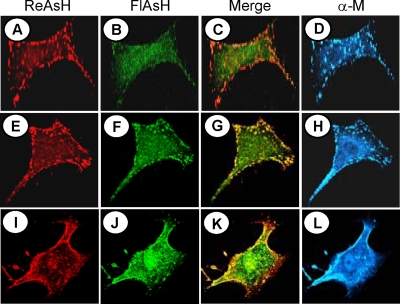
FIG. 6.
Dynamic imaging of existing and newly synthesized pools of M protein by dual biarsenical labeling. BHK-21 cells infected with VSV-ΔM-Mtc were treated with ReAsH for 30 min at 4 hpi (A, E, and I), washed, and then labeled immediately (A, B, C, and D) or 1 h (E, F, G, and H) or 2 h (I, J, K, and L) later with FlAsH for 30 min (B, F, and J). Following biarsenical dye labeling, the cells were washed, fixed, immunostained with anti-M MAb and Alexa 350-conjugated secondary antibody (D, H, and L), and examined by confocal fluorescence microscopy as described in Materials and Methods. Merged images of cells stained with ReAsH and FlAsH are shown in panels C, G, and K.

