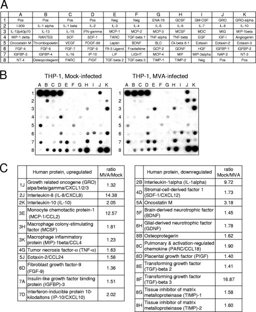
FIG. 2.
Detection of differentially expressed proteins in supernatants of MVA-infected THP-1 cells. (A) Scheme of the spotted primary antibodies on the RayBio human cytokine antibody array V. (B) Images after chemiluminescence detection of supernatants from MVA-infected (MOI of 4) and mock-infected THP-1 cells, which were incubated in VLE-RPMI 1640/0.5% FCS for 16 h. (C) List of differentially regulated proteins. The ratios indicated are calculated by using the intensities of the corresponding protein spots after background (Neg) correction and normalization of the intensities according to the mean intensities of the positive controls (Pos).

