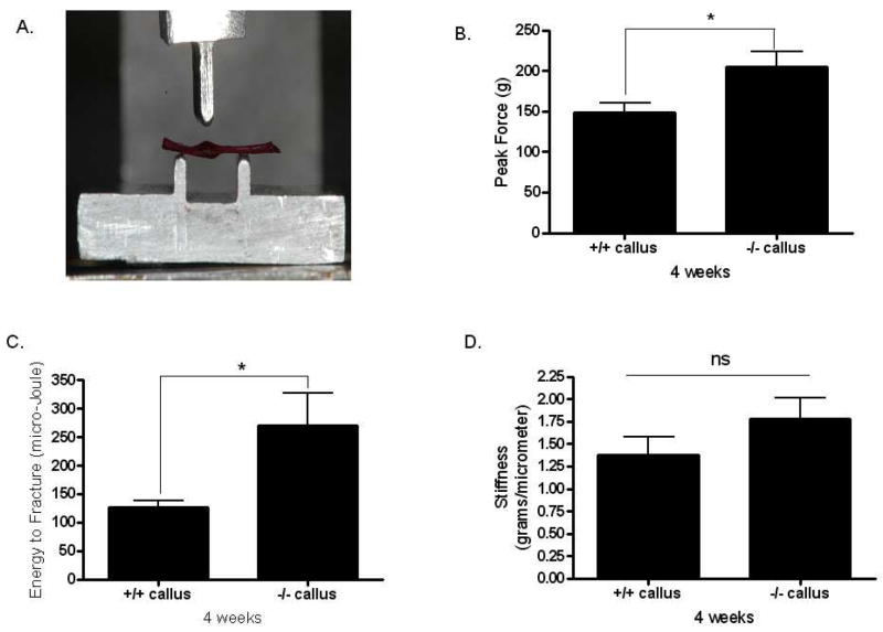
FIG. 5.
Biomechanical data from fracture calluses four weeks post-surgery. (A) Alizarin-red stained fibula specimen showing position of the ossified callus in the three-point bending platform. (B) Ultimate force (peak load) and (C) energy to fracture (toughness) are significantly increased in Mstn−/− mice, and stiffness (D) is non-significantly increased in Mstn−/− mice. * p < 0.05.

