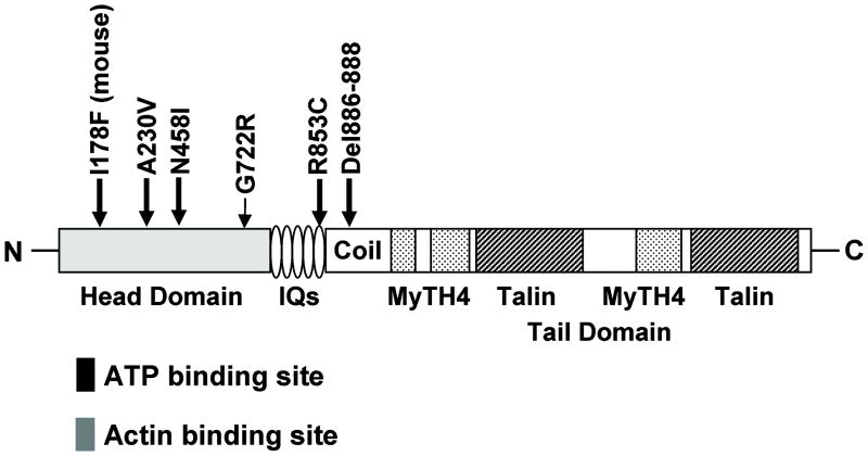
FIG. 1.
Dominant mutations within Myosin VIIA. Schematic representation of MYO7A functional regions indicates the location of five DFNA11 mutations and the dominant Hdb mouse mutant. The head region (light gray shading) contains ATP (black box) and actin (dark gray box) binding sites. The tail domain contains four notable regions: 1) IQs represent five light-chain-binding repeats; 2) coil indicates coiled-coil domain that may be involved in dimerization; 3) MyTH4 indicates myosin tail homology-4 domains that are regions conserved between myosins; and 4) talin represents talin-like homology domains which are predicted to bind actin.

