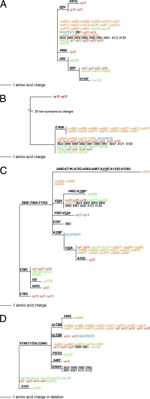
FIG. 3.
Protein phylograms of mdh (A), fumC (B), tonB (C) and fimH (D) genes of K. pneumoniae derived from the corresponding DNA trees by collapsing the branches carrying only synonymous variations. Structural mutations are shown along the branches. Structural mutations [or L(−12) deletion in panel A for FimH] that occurred multiple times across independent lineages of DNA phylogeny are termed hot-spot mutations and are shown as two groups (as in Fig. 1D). Different colors are used for strains from different origins of isolation: orange for UTI, red for sepsis, green for environment, black for liver, and blue for sputum.

