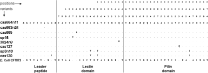
FIG. 4.
Amino acid polymorphisms in the FimH proteins of K. pneumoniae structural variants and E. coli CFT073, showing domain borders of the E. coli FimH protein, which are as follows: in amino acid positions 1 to 21, the leader peptide; in positions 22 to 177, the lectin domain; in positions 178 to 180, the linker chain; and in positions 181 to 300, the pilin domain. The dots correspond to identical amino acids relative to the consensus FimH of K. pneumoniae (represented by cas664). The numbers above the sequences indicate corresponding positions of the structural mutations. The numbers in parentheses correspond to the number of isolates carrying the FimH variant.

