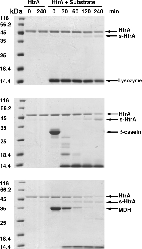
FIG. 1.
HtrA autocleavage in the presence of substrate. Proteolytic reactions were assembled in a total volume of 250 μl containing 1.3 μM HtrA and 13 μM concentrations of substrate. The substrates tested were lysozyme (top panel), β-casein (middle panel), and MDH (bottom panel) (lanes labeled “HtrA + Substrate”). A control reaction was performed for each degradation assay consisting of HtrA protein incubated in the absence of substrate (lanes labeled “HtrA”). Reactions were incubated and, at the indicated time points, samples were obtained, and the products of the reaction resolved by SDS-PAGE and stained by Coomassie brilliant blue. Arrows indicate the bands for the corresponding substrate, full-length HtrA and s-HtrA protein in each gel.

