
Fig. 5.
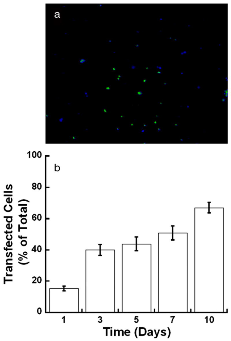
Fluorescence images of MCF-7/WS8 cells at day 7 encapsulated within an 8% PEG-Acryl/1% HA-Acryl hydrogel and transfected with PEI/pGFP-LUC complexes (a). Cells expressing GFP appear green while cells stained with Hoechst appear blue. Quantification of the percentage of transfected cells (b).