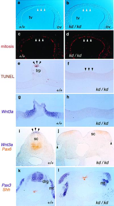
Figure 3.
Development of the roof plate is disturbed in the Zic2kd/kd embryo. Immunohistochemistry (a–d), TUNEL staining (e and f), and in situ hybridization (g–l) were performed on Zic2+/+ (a, c, e, g, i, and k) and Zic2kd/kd (b, d, f, h, j, and l). (a–d) Immunohistochemical staining using antiphospho-histone H3 antibody to detect mitotic cells in the sections through E9.5 telencephalon. (a and b) Bright-field view. In the Zic2+/+ animal, the mitotic cells were scarce in the prospective roof plate region with thinning (arrowheads in a and c). Such a scarcity or thinning was not observed in the Zic2kd/kd animals (arrowheads in b and d). (e and f) TUNEL staining of the sections through the E10.5 telencephalic roof plate (trp). In the Zic2+/+ animal, staining characteristic of dying cells was observed at the midline (e, arrowheads) whereas no staining in the corresponding region of Zic2kd/kd animals was observed (f, arrowheads). (g–l) In situ hybridization showing the distribution of Wnt3a (g, h, i, and j, purple), Pax6 (i and j, orange), Pax3 (k and l, purple), and Shh (k and l, orange), in transverse sections through telencephalic roof plate (g and h) and through the lumbar spinal cord of E10.5 embryos (i–l). Note that Wnt3a expression is absent in the telencephalic roof plate (h) and reduced in the edges of the open spinal cord (j, arrowheads), which corresponds to the roof plate of the properly closed spinal cord (i, arrowheads). Pax3 staining in the dorsal root ganglia (drg) of Zic2kd/kd embryos was hardly visible whereas spinal cord (sc) and myotome (mt) staining remained visibly unchanged (k and l).

