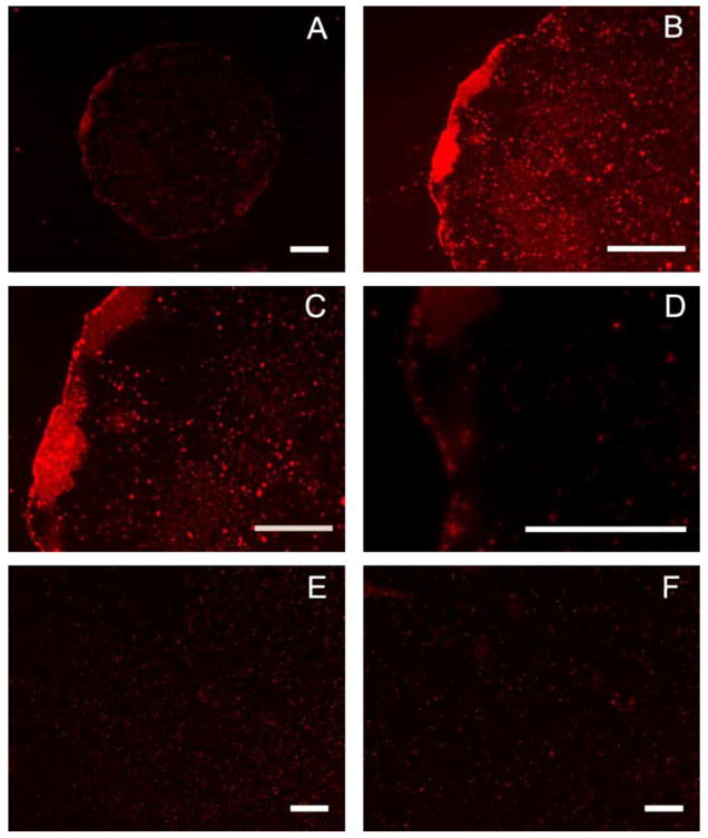
Fig. 7.
Patterned complex deposition. Condensation figures were utilized to confine droplets containing rhodamine-labeled DNA complexes to hydrophilic regions of a patterned surface, resulting in patterned DNA immobilization (A–D). Complexes were allowed to deposit for 1 h, in humid conditions, and then visualized with fluorescence microscopy. Control complex deposition was performed on unpatterned 50% MUA (E) and DT10 (F) SAMs. Scale bars correspond to 200 μm (A, B, E, F) and 100 μm (C, D).

